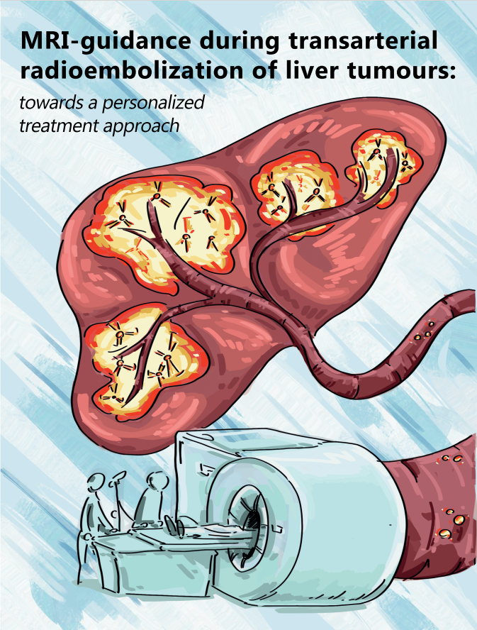
MRI-guidance during transarterial radioembolization: towards a personalized treatment approach
Transarterial radioembolization (TARE), also called selective internal radiation therapy (SIRT), is a treatment modality for liver tumours during which radioactive microspheres are injected into (subbranches of) the hepatic artery through a catheter. These microspheres are distributed in the perfused liver volume, both in tumours and healthy liver tissue, as a result of the blood flow. In order to achieve the optimal treatment efficacy, an adequate tumour dose should be achieved, combined with an as low as possible healthy tissue dose. In recent clinical studies, it has been demonstrated that an increased treatment personalization can improve treatment efficacy and therefore improve patient outcomes. The holmium-166 (166Ho) microspheres used for TARE in this thesis are unique with respect to their imaging possibilities, specifically the ability to visualize and quantify the microspheres with MRI.
The overarching aim of this thesis was to develop an image-guided approach to 166Ho TARE, in which the clinician can assess the achieved dose distribution during the procedure in semi real-time using MRI. This way, treatment parameters can potentially be adjusted during the procedure, in order to improve the dose distribution if for instance tumours or parts of tumours are undertreated.